Intensive visual development in children
Intensive visual development lasts until the end of the 7th year of life. This is a key period for treating eye diseases in order to ensure the normal development of the child’s eyes and vision.

When should you take your child for an eye exam?
An ophthalmological examination of a child is one of the most important examinations and should be done by the age of 2. In prematurely born children, the first examination should be done by the age of 6 months. If there is a positive family history of amblyopia and if eye deviation is noticed, the first examination should be done by the age of 1.
The next mandatory preventive ophthalmological examination should be done when the child turns 4 and before starting school.
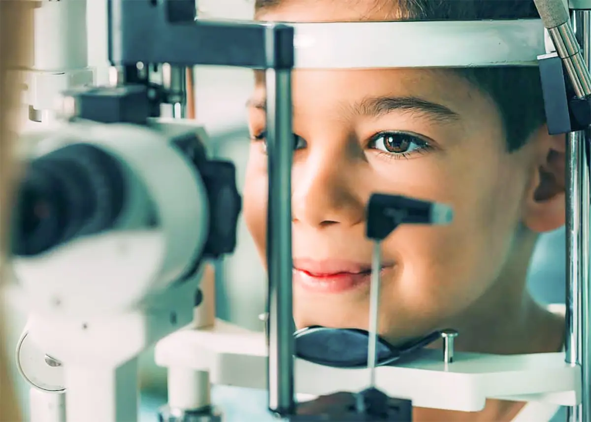
What does a pediatric eye examination look like?
An ophthalmological examination of a child lasts up to 2 hours and includes:
Determining visual acuity for near and distance
In very young babies, special pattern cards are used to determine which pattern size captures the baby’s attention, and the result is then compared with charts prescribed for that age.
For preschool children, pictures (Lea symbols) are used, which are made according to the same standards as letters for adults. For the best possible insight into visual acuity, it is necessary to examine how the child recognizes pictures both at near and at distance. For school-age children, letter charts are used, as for adults.
Tests for strabismus and stereovision
Taking the orthoptic status serves to check the position and proper mobility, as well as the cooperation between both eyes. In this part of the examination, the type and size of strabismus are determined, disorders of binocular vision, amblyopia (lazy eye), and nystagmus (eye oscillation) are identified.
The development of stereovision (3D vision) is also assessed with the help of stereotests (Titmus test, Lang test). In children or adults with nystagmus, the necessary additional measurements are performed in this part of the examination.
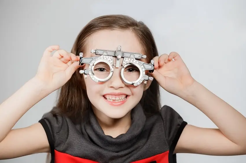
Pupil dilation and determination of objective refraction
Due to the strong accommodative ability of the child’s eye, which can sometimes easily mask even high refractive errors, it is necessary to dilate the pupils by instilling drops several times (a total of 1 hour).
It is advisable to prepare the child at home for the fact that drops will be put into the eyes and that vision will be blurred after the examination (from several hours up to a day).
After the pupils are fully dilated, objective refraction is determined – skiascopy. This is important for detecting latent refractive errors or other visual disorders and for prescribing appropriate glasses. Objective refraction can be determined even in the youngest children because their cooperation is not required.
Complete examination of the anterior and posterior segment of the eye
The most common eye diseases in children
Strabismus (crossed eyes) is a disorder of eye position or motility in which both eyes do not share a common direction of gaze or the mobility of one eye is limited. In children, strabismus is most often congenital or develops in early childhood, but it can also occur due to injuries and various diseases. Most often the eye turns inward (esotropia) or outward (exotropia), and more rarely vertically. It is often associated with other visual disorders.
Treatment depends on the type of strabismus, its severity and the child’s age: appropriate correction of refractive error, patching (occlusion) of the better eye, or surgery.
Cataract in children can be congenital. If it is not removed early, vision will not develop. Urgent surgery is required.
Eye tearing can be a consequence of infection or failure of the channel between the eye and the nose to open. It is most often resolved spontaneously by the age of 1.
- In 90% of children, the duct opens spontaneously during the first year of life.
- In a smaller number of children, a probing procedure under general anesthesia is required.
- In rare cases, a silicone tube is inserted and remains in place for 6 months.
If vision is reduced in only one eye, the child unconsciously uses the better eye while neglecting the weaker one. This leads to the development of amblyopia (lazy eye).
If this happens at an early age, vision may remain permanently reduced. The risk is higher if there is a similar problem in the family.
Treatment of amblyopia (occlusion)
When an amblyopic eye is detected, the child needs to be prescribed glasses and occlusion (patching) of the healthy eye must be carried out. The occlusion period varies from 1–6 hours a day. The patch (occluder) is placed over the eye, not on the glasses. The effect is greater if the child draws, reads or plays during this time.
Children often resist wearing a patch, especially if they see poorly with the weaker eye. Patience and persistence are needed to achieve the desired effect.
The earlier it is detected, the greater the success of treatment. If left untreated, vision will never regain good quality, which later also affects the choice of profession.
Certificates of bravery