
Meibomian gland expression – the key to the health of your tear film and clear vision
What are Meibomian glands? Meibomian glands are small glands located in the eyelids that produce the lipid (fatty) component of…

The disease glaucoma or green cataract, as it is popularly known, primarily refers to damage to the optic nerve. The Optic nerve is a structure located on the back segment of the eye, i.e. on the fundus (picture 1).
However, the mechanism of glaucoma itself is most often associated with structures located in the front third of the eyeball and are part of the anterior and posterior chambers of the eye. Analysis of the structures that make up the anterior chamber of the eye is of great importance in diagnosing patients with glaucoma, as well as in deciding on the type of therapy to be applied.
Pentacam HR, which our clinic owns, represents the third generation of Pentacam devices and is the newest type of device of its kind in the world (Figure 2). This device enables fast, painless, non-contact recording of the morphological features of the anterior chamber. Recording with this device takes only two seconds, which results in at least 50 photos (picture 3), which are automatically reconstructed into a 3d photo (picture 4), which gives us an insight into the following important data:
[nggallery id=23]
Perfect Vision is an Eye Laser Surgery Center located within the Kuća zdravlja Polyclinic, headquartered in Subotica.
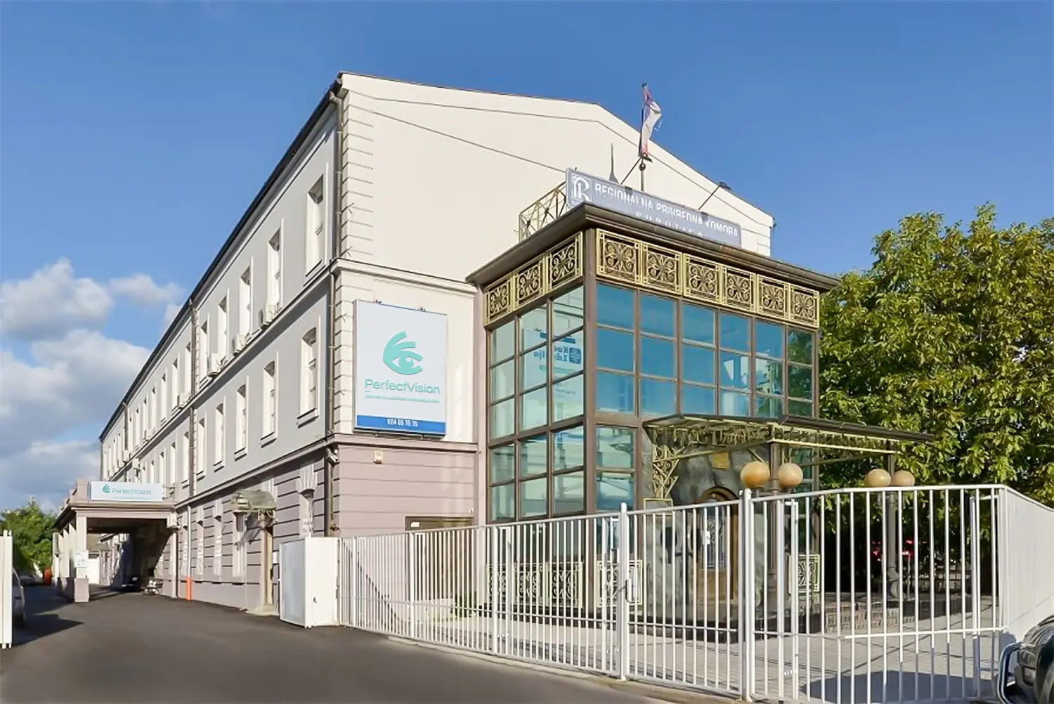
Perfect Vision is a Center for Laser Eye Surgery, located within the Polyclinic House of Health, based in Subotica. Visit us!
We have an ISO certificate according to ISO 9001: 2015, registration number 12 100 59137 TMS for the area of Diagnostic, Conservative and Surgical Ophthalmic Services, issued by the certification body TÜV NORD CERT GmbH.